1.1 Revision of Autonomic Nervous System (ANS)
John Smithson
Revision of Autonomic Nervous System
In this topic, we will revisit the autonomic nervous system (ANS) taking a look at the structures within it. We will look at the different types of neurotransmitters and the main receptors that are involved with neuromodulation of the ANS and the target response.
Be able to:
- describe the relationship between the autonomic nervous system and the central nervous system.
- describe the function of the autonomic nervous system.
- describe the structural arrangement of the autonomic nervous system.
- relate the activity of the key neurotransmitters on the main receptors in the autonomic nervous system and the target organ response.
The Central Nervous system (CNS)
To understand its role, let’s look at the CNS in a broader context.
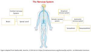
The CNS consists of the brain and spinal cord, and the peripheral nervous system consists of nerves that run to the rest of the body.
- The peripheral nervous system (PNS) has both afferent and efferent function and is divided into the somatic and autonomic systems.
- The somatic nervous system carries signals from the body to the central nervous system as well as carry motor signal from the CNS to the skeletal muscles. It is the somatic nervous system that is allowing me to type this text right at this moment using my fingers on the keyboard, and now reaching for my coffee. The movement is voluntary.
- The second division of the PNS is the autonomic nervous system. The autonomic nervous system (ANS), is responsible for conveying all outputs from the CNS to the body, excluding muscle innervation. It relays signals from the CNS to the internal organs and from the internal organs to the CNS.
- The autonomic nervous system (ANS) is a crucial part of the central nervous system (CNS) and serves as one of the two main control links between the CNS and the rest of the body. The ANS operates largely involuntarily and comprises two main subdivisions: the sympathetic and parasympathetic systems. There is also a third component, the enteric nervous system, which affects the gastrointestinal system but is less relevant to this topic.
The primary functions of the ANS include regulating:
- Smooth muscle contraction and relaxation in the bladder, uterus, gastrointestinal tract, and the walls of blood vessels.
- All exocrine functions (glands that secrete substances into ducts, such as sweat, tears, mucous, and saliva) and some endocrine functions (such as the release of hormones like adrenaline from the adrenal glands).
- Heart rate and contractility.
- Energy metabolism in the liver and skeletal muscle.
Figure 1.1.1 illustrates the nervous system and the divisions of the somatic and autonomic nervous system within the peripheral nervous system.
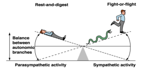
The sympathetic and parasympathetic nervous systems are sometimes referred to as the “fight and flight” (sympathetic) system and the “rest and digest” (parasympathetic) system. Some use the rough differentiator of Energy spending (sympathetic) and energy conservation (parasympathetic). They are a simplification of a complex system so let’s illustrate these systems starting with the activation of the sympathetic nervous system. The sympathetic nervous system uses catecholamines (mostly noradrenaline and some adrenaline) to increase heart rate and blood pressure, dilate blood vessels and bronchioles, and dilate pupils. Useful to the guy running from the snake in Figure 3 below (or you before your next OSCE).
In contrast, the parasympathetic nervous system decreases heart rate and contractility, constricts the bronchi, increases gastrointestinal motility, and constricts the pupils. While this might suggest they have opposite and opposing effects, it is an oversimplification of a complex system.
Some target organs are regulated by only one of the two systems. For example, the vasculature responds only to sympathetic signals, with vasoconstriction resulting from the release of other neurotransmitters. Additionally, some target organs respond similarly to both sympathetic and parasympathetic stimulation. For instance, salivary glands alter salivary secretion in response to both systems.
Knowledge check
Innervation of the somatic nervous system
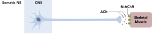
The sensory and motor neurons of the sensory-somatic system have a single synapse between the organ and a central nervous system (CNS) neuron. These synapses use acetylcholine, which interacts with nicotinic acetylcholine receptors to transmit signals across the gap.
Innervation of the autonomic nervous system
In contrast, the autonomic nervous system communicates with its target organs using neurons arranged in series. The autonomic nervous system involves two types of neurons:
- Shorter preganglionic neurons
- Longer postganglionic neurons.
In the autonomic nervous system, all short preganglionic neurotransmitters are acetylcholine (ACh), which interacts with nicotinic receptors (N-Ach-R). This changes in the postganglionic neurons. The neurotransmitter released by the longer postganglionic neuron is either noradrenaline (NA) (and sometimes adrenaline if the adrenal medulla is stimulated) in the sympathetic nervous system or acetylcholine if the parasympathetic nervous system is activated, depending on the target organ and the branch of the autonomic nervous system.
🔑 Key Concept: Autonomic nervous system function
The autonomic nervous system is divided into the parasympathetic and sympathetic nervous system. The signal from the CNS first travels along the pre-ganglionic neuron terminating in the ganglion where it releases acetylcholine which interacts with N-AChR receptors. The signal is passed to the post-ganglionic neuron where it reaches the target organ (or very close to) and releases either acetylcholine (in the case of parasympathetic nervous system activation) where local muscarinic-acetylcholine receptors (M-AChR) are stimulated or where the sympathetic nervous system is activated, noradrenaline from the post-ganglionic neuron or adrenaline (from the adrenal medulla) is released. Both noradrenaline and adrenaline interact with adrenergic receptors – alpha or beta receptors.
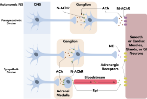
So, as you know, the postganglionic parasympathetic neuron releases acetylcholine and terminates very close to the target organ, making it short compared to the sympathetic postganglionic neuron. Acetylcholine released from the postganglionic parasympathetic nerve interacts with muscarinic receptors. The longer postganglionic sympathetic neurons release noradrenaline, which interacts with either α or β receptors.
Pharmacology basics refresh
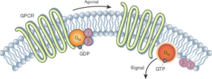
The adrenoreceptors are G-protein-coupled receptors. G-protein-coupled receptors (GPCRs) are a large family of cell surface receptors that play a crucial role in signal transduction, allowing cells to respond to external stimuli. When a ligand, such as the neurotransmitter noradrenaline, binds to a GPCR, it activates an associated G-protein by causing the exchange of GDP for GTP on the alpha subunit of the G-protein. This activation triggers a cascade of intracellular signalling pathways, influencing various cellular responses such as metabolism, growth, and differentiation. GPCRs are involved in numerous physiological processes and are significant targets for a wide range of therapeutic drugs.
📺 Watch vodcast: The autonomic nervous system (2m 56s)
Note: in the text above I align post-ganglionic release of noradrenaline and adrenaline with the sympathetic nervous system and the release of acetylcholine with the parasympathetic nervous system. There are a few exceptions in the SNS where the post-ganglionic neuron release acetylcholine as the primary neurotransmitter innervating the sweat glands and some blood vessels. For simplicity sake, it is generally safe to allocate post ganglionic acetylcholine release with the parasympathetic NS and adrenaline/noradrenaline with the sympathetic nervous system.
🔑 Key concept
Knowledge check
Neuromodulation of the ANS: Neurotransmitter release (either acetylcholine from the parasympathetic postganglionic neuron or noradrenaline or acetylcholine from the postganglionic sympathetic neuron) release their respective neurotransmitters from the neuron in response to electrical activity of the nerve fibre. As the nerve depolarises, calcium enters the nerve cell causing the expulsion of vesicles containing the neurotransmitter (ACh or NA for example). The neurotransmitter release interacts with the target organ to exert its effect. It also may interact with other neurons to alter the release of neurotransmitter. For example, the release of noradrenaline or adrenaline from a sympathetic postganglionic neuron may inhibit the local release of acetylcholine from the parasympathetic postganglionic neuron. This means the opposing effects are achieved not only because of the effect of noradrenaline at the target organ but also because of the absence of an opposing effect of acetylcholine. This is described as a heterotropic interaction. Homotropic interactions, where the released neurotransmitter acts as an auto-regulatory mechanism (inhibiting further release of the neurotransmitter from the same terminal can also occur as a means of negative feedback.) Postsynaptic modulation of the ANS can occur where neurotransmitters interact with postsynaptic structures (neurons, cells etc.) to alter their response.
Terminating neurotransmitter action. Once the neurotransmitters are released, there needs to be a mechanism to limit the duration and location of the effect of the neurotransmitter. Think of these interactions as a very short-term temporary stimulation. The body needs to the signal on and then have it turn off. To have this quick on-off cycle, there are two main mechanisms that cause the breakdown of the neurotransmitter or reuptake of the neurotransmitter from the synaptic cleft into the presynaptic terminal.
Acetylcholine in the synaptic cleft is rapidly broken down to an inactive form by the enzyme acetylcholinesterase (AChE). This enzyme is largely responsible for the removal of the neurotransmitter and thus terminating its effect. In contrast, noradrenaline is transported from the synaptic cleft into the presynaptic neuron by active transport by noradrenaline transporter (NET).
📺 Watch vodcast: Termination of ANS neurotransmitter action video 2 (1:44s)
Concept check
Summary
- The ANS is made up of the Sympathetic Nervous System and the Parasympathetic Nervous System.
- The SNS and PNS are made up of the preganglionic neuron and postganglionic neuron in series. The preganglionic neuron. releases acetylcholine and this Ach interacts with nicotinic receptors in the postganglionic neuron.
- Depending on the post ganglionic neuron it either releases
- Noradrenaline which interacts with either α or β receptors (sympathetic subdivision).
- Acetylcholine with interacts with muscarinic receptors sympathetic and parasympathetic subdivisions).
- The release and subsequent effect of the release neurotransmitters from the PNS and SNS can be modulated by either homotropic or heterotropic means.
- The effect of released neurotransmitters is kept short and local by two main mechanisms – breakdown of acetylcholine by acetylcholinesterase or reuptake of noradrenaline by NET.
COMMONWEALTH OF AUSTRALIA Copyright Regulations 1969 WARNING
This material has been reproduced and communicated to you by or on behalf of James Cook University in accordance with section 113P of the Copyright Act 1969 (Act).
The material in this communication may be subject to copyright under the Act. Any further reproduction or communication of this material by you may be the subject of copyright protection under the Act. Do not remove this notice.